Overview
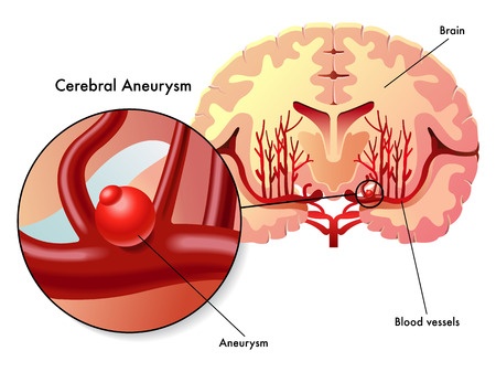
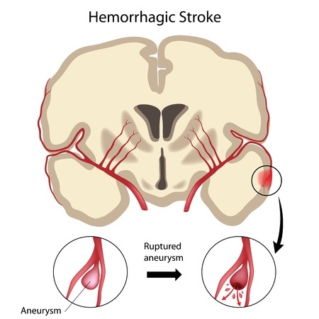
A brain aneurysm is a section of blood vessel in the brain that has bulged or ballooned. This section of blood vessel actually looks like a berry hanging on a vine. Brain aneurysms are dangerous because they can burst and cause serious bleeding within the brain called hemorrhagic stroke. Ruptured brain aneurysms prove fatal in thirty percent of cases. It is also possible for brain aneurysms to leak as oppose to completely bursting. However, in most cases, brain aneurysms do not burst, yet they cause a host of symptoms and medical issues. Nevertheless, brain aneurysms that do not burst can be treated to prevent them from bursting later.
Symptoms
Since brain aneurysms fall into the categories of ruptured, leaking, and unruptured, you may experience different symptoms depending on which category of brain aneurysm you have.If you have a ruptured brain aneurysm, then you will most likely suffer from a sudden, severe headache that many patients describe as the worst headache they have ever experienced. Other symptoms of ruptured brain aneurysms include seizure, loss of consciousness, confusion, drooping eyelid, blurred or double vision, sensitivity to light, stiff neck, nausea, or vomiting.
Leaking aneurysms also produce a sudden and severe headache. Although leaking aneurysms almost always become ruptured aneurysms, a severe headache is the only symptom associated with leaking brain aneurysms.
Commonly, unruptured brain aneurysms do not produce any symptoms. But, unruptured brain aneurysms sometimes grow so large that they press on nearby brain tissues and nerves. This pressure may cause symptoms like pain behind the eye, a change in vision, a drooping eyelid, or dilated pupils. You could also experience numbness, weakness, or paralysis on one side of your face.
Risk Factors
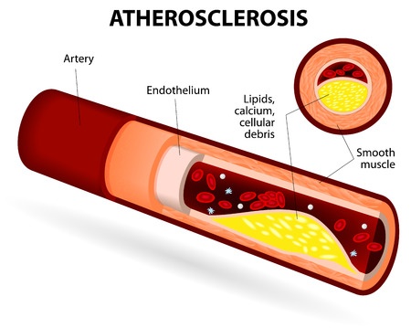
Brain aneurysms affect more adults than children and more women than men. In addition, those affected by brain aneurysms possess other risk factors such as hardened arteries referred to as atherosclerosis, high blood pressure, blood infections, low estrogen levels, and old age. Smoking, head injuries, drug abuse, and excessive alcohol consumption also increase your chances of developing a brain aneurysm.Some heredity factors and birth defects can put you at a greater risk of getting a brain aneurysm. Connective tissue disorders which weaken blood vessels, polycystic kidney disease which increases blood pressure, and a family history of brain aneurysms all represent examples of hereditary factors. On the other hand, birth defects include an abnormally narrowed aorta and an abnormal connection between the arteries and veins of the brain called cerebral arteriovenous malformation.
Diagnosis
If you experience any of the symptoms connected to brain aneurysms, you should seek medical attention immediately. Your doctor will conduct one or more diagnostic tests to examine your brain and its surrounding tissues for signs of bleeding and stroke. These tests include computerized tomography (CT), a cerebrospinal fluid test, magnetic resonance imaging (MRI), and a cerebral angiogram.
- Computerized Tomography (CT) - also known as CT angiography, is a special X-ray that yields images of your entire brain in two-dimensional slices. Your doctor may inject dye intravenously to increase the visibility of blood flow through your brain. Observing how well the dye travels through your brain can help your doctor determine if you have an aneurysm.
- Cerebrospinal Fluid Test - tests the fluid surrounding your brain and spine called cerebrospinal fluid, or CSF, to check for the presence of red blood cells. The presence of red blood cells in the CSF is often a good indicator of bleeding in the brain. This process is called a spinal tap and is only conducted if a CT scan does not reveal any evidence of bleeding.
- Magnetic Resonance Imaging (MRI) - or MRI angiography, uses a magnetic field and radio waves to produce two-dimensional slices or three-dimensional images of your brain. Introducing a dye intravenously will further assist your doctor in visualizing blood flow through the vessels of your brain as well as locate your ruputured aneurysm. This test often produces clearer results than a CT scan.
- Cerebral Angiogram - also called a cerebral arteriogram, this test involves your doctor inserting a thin, flexible tube into a large artery in your body and guiding it up past your heart into the arteries of your brain. He or she will then inject a special dye into the tube which will travel throughout the arteries of your brain. Finally, a series of X-rays will reveal how well the dye traveled through your brain and ultimately locate the site of your ruptured aneurysm. Considering the invasiveness of this process, this test is usually reserved as a last resort for diagnosis.
Treatment
If you are diagnosed with a ruptured brain aneurysm, then your doctor will quickly refer you to a neurosurgeon that will pursue one of two treatments. The primary goal of both of these treatments is to stop blood flow to the aneurysm. Both treatments pose risks like re-bleeding or loss of blood flow to the brain, yet your neurosurgeon will take these risks into account before treating your aneurysm.The first treatment, surgical clipping, closes the ruptured aneurysm. Your neurosurgeon achieves this by removing a section of your skull and finding a blood vessel that feeds the section of blood vessel where your aneurysm is located. Next, he or she will fasten a small metal clip at the base of the aneurysm to cease blow flow to it.
The second treatment, endovascular coiling, is less invasive than surgical clipping and similar to a cerebral angiogram. In endovascular coiling, your neurosurgeon inserts a hollow, plastic tube into a large artery and guides it up past your heart into your brain right to the site of your aneurysm. Then, he or she will use a guide wire to push a soft, platinum wire through the tube and into your aneurysm. That platinum wire coils up inside of the aneurysm, interrupts blood flow, and develops a blood clot within the aneurysm. This blood clot ultimately seals off the aneurysm from the artery that supplies it.
Other treatments available for treating ruptured brain aneurysms include anti-seizure medications, calcium channel blockers, stroke-preventing interventions, pain relievers, rehabilitative therapy, and ventricular or lumbar draining.
How surgeons treat unruptured brain aneurysms depends heavily on the size and location of your aneurysm as well as your age, family history, and overall health. Surgical clipping and endovascular coiling are sometimes employed to prevent the rupture of an unruptured brain aneurysm. In other situations, managing your blood pressure with diet, exercise, or medication is sufficient to lower your risk of rupture.
In the end, having the right surgeon can dramatically decrease your risk and D.R.A. Medical specialists have extensive experience in treating brain aneurysms.